World Brain Tumor Dayn 2026: A Closer Look.
Every 8th of June World Brain Tumor day attempts to inform about brain tumors across the world. In 2000 the German Brain Tumor Association began this campaign that is now International. The aims of this day are to increase awareness of brain tumors and their implications, to encourage an earlier and accurate diagnosis, to encourage an earlier and accurate diagnosis, and the support of patients and to make further funds available and the support of patients and to make further funds available for the development of improved therapies. Brain tumors – type and consequences.
The World Brain Tumor Day 2026 theme
The World Brain Tumor Day 2026 theme “A Closer Look” encourages everyone — from medical professionals to the general public — to pay closer attention to the early warning signs of brain tumors and to advocate for improved diagnostic capabilities worldwide. This theme is especially relevant as early detection remains one of the most critical factors in improving patient outcomes.
This theme is especially relevant as early detection remains one of the most critical factors in improving patient outcomes.
Brain Tumors – Primary or Secondary?
When brain cells start to grow abnormally and accumulate into a mass inside the brain, it is called a brain tumor. The brain cells themselves are the source of a primary brain tumor. These types of brain tumors can arise from glial cells (astrocytes, oligodendrocytes, and ependymal cells), neurons, and other specialized cells like those found in the meninges (meningiomas), the nerve sheet cells (neurinomas), choroid plexus, and embryonal tissues.
When a brain tumor is a metastasis from body cancer cells that have entered the brain through the bloodstream, it is referred to as “secondary.” Brain metastases are the most frequent cause of malignant tumor of the central nervous system (brain and spinal cord), four-times higher than primary tumors. Cancer types that most frequently metastasize to brain originate in the lung (≥50%), breast (15–25%), or skin (melanoma, 5–20%).
Brain Tumors – Benign or Malignant?
Certain primary brain tumor types, such as glioblastoma, astrocytoma, or oligodendroglioma, have the potential to spread into the surrounding brain tissue. The tumor cells disturb the normal function of the normal brain cells. In addition, their invasive character makes it difficult to remove them by surgery in total.
Many other brain tumor types such as meningiomas, or pituitary tumors do not invade the brain but squeeze and displace brain tissue. The enclosed space inside the skull is limited. Therefore, even when benign tumors grow, they start closing in the space and provoke irritation and damage to the brain.
Therefore, whether invasive or not, whether malignant or benign, many of these brain tumor types will cause neurological symptoms and can be life-threatening and life-limiting.
Brain tumors can occur in more than hundred types, each with a unique range of symptoms, treatment requirements, and prognoses Brain tumors can have profound effects on a patient’s physical, mental, and emotional well-being.
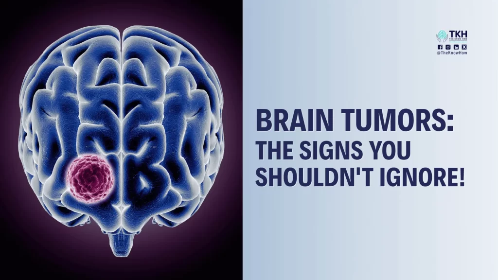
Brain Tumor Symptoms – Get Alert!
Clinical signs of brain tumors are highly variable based on the location, size, and type of the tumor. Most of the symptoms are unspecific and can also be caused by other conditions.
A comprehensive neurological symptom assessment can help determine whether these signs are linked to a brain tumor or another underlying condition.
Nevertheless, when you or your loved ones become aware of symptoms listed below, talk to your doctor about it.
Brain tumor symptoms may include:
- severe headaches,
- numbness and tingling in an arm or leg,
- memory or thinking problems,
- nausea and vomiting,
- changes in speech, vision or hearing,
- seizures,
- changes in personality.
Symptoms may start gradually over weeks to months or rapidly, potentially leading to an emergency visit at the hospital.
Who Diagnoses a Brain Tumor?
The Neuroradiologist: Look into the Brain
When a patient exhibits symptoms of nerve and brain dysfunction, a neurologist will do a physical neurological examination first. If the results point to an origin of symptoms within the brain, diagnostic imaging such as a CT scan (computerized tomography) or an MRI scan (magnetic resonance imaging) will be performed.
Advanced brain imaging and diagnostic assessment can provide critical information for identifying abnormalities and guiding the next stage of care.
These tests allow the neuroradiologist to “see” your brain to identify if there is a tumor. If so, it is important to determine where it is located, if it is demarcated off from brain tissue, and its size. You may then be referred to a neurosurgeon, who, if necessary, will recommend a biopsy or surgery.
The Neuropathologist: Making Exact Diagnosis
Biopsy or surgery are required to ascertain the origin and nature of the tumor. Is it primary or metastatic, from which cell types does it arise, is it growing slowly or aggressive? What other characteristics do the tumor cells display?
The diagnostic activities of a neuropathologist include intraoperative consultations, histopathological evaluation, and the use of specific immunological markers. In addition, according to the WHO 2021 classification of brain tumors, molecular genetic examinations have become essential for the exact diagnosis. Molecular genetics also give important information on the prognosis and help to predict the benefit of therapeutical interventions on an individual basis.
Molecular profiling and personalized treatment planning may help physicians identify therapies that are better aligned with an individual’s tumor characteristics.
Treatment of Brain Tumors – It´s Teamwork
in recent years, there have been several advancements in the way brain tumors are diagnosed and treated. These innovations aim to make treatments more precise, reduce side effects, and improve overall outcomes. Here are the most promising developments:
Image-Guided Surgery
Image-guided surgery: Modern imaging techniques now allow surgeons to view the tumour in great detail during surgery. Tools like intraoperative MRI and neuronavigation systems support safer and more effective procedures.
Before proceeding with complex brain tumor surgery, obtaining an independent review of surgical recommendations may help patients better understand their available options.
Proton Therapy
Proton therapy: uses proton beams instead of traditional X-rays, directed more precisely to the tumour, protecting nearby healthy tissues. Especially useful for brain tumours in children.
Targeted Drug Therapy
Targeted drug therapy: targeted drugs act specifically on identified genetic changes in brain tumours, working differently from standard chemotherapy with potentially fewer side effects.
Advances in precision oncology are making it possible to match treatments to specific genetic alterations identified within a tumor
Tumour Treating Fields (TTF)Tumour Treating Fields (TTF)
Tumor Treating Field (TTF): This non-invasive wearable device uses low-intensity electric fields to interfere with cancer cell division and slow tumour growth, used alongside chemotherapy for glioblastoma.
Liquid Biopsy
Liquid Biopsy: Liquid biopsy detects tumour DNA in blood or spinal fluid, offering a less invasive way to monitor the tumour’s progress and treatment effectiveness without surgery.
Biomarker-based tumor monitoring can provide valuable insights into disease progression and treatment response over time.
Other ways of treatments:
The Neurosurgeon: Surgical Removement of brain tumors
Surgery is the most common first line treatment for brain tumors,and often it may be the only treatment needed.
Patients considering brain tumor surgery may benefit from a specialist second opinion to better understand potential risks, benefits, and alternatives.
There are numerous approaches neurosurgeons take to remove brain tumors depending on their size and location.
In a craniotomy a piece of skull is temporarily removal to allow surgeons access to the brain. Neurosurgeons may also enter the brain through other parts of the body, like the nose, to better reach certain regions and minimize scarring. If the tumor is in a challenging position in the brain, a biopsy might be the initial choice to clarify its type by the neuropathologist.
Surgery is rarely the only path of treatment for tumors that are growing aggressively. In these situations, the treating team is typically completed by a radiation oncologist and a neuro-oncologist, who communicate with the patient and their family in deciding the optimal course of care.
The Radiation Oncologist: High-Energy Radiation Kill Brain Tumor Cells
Radiotherapy for brain tumors is used to destroy cancer cells, shrink tumors, and relieve symptoms. It uses high-energy radiation to damage the DNA of tumor cells, preventing them from growing and dividing.
Radiotherapy is differentiated by how the radiation is applied: from outside the body or directly from within the tumor.
External beam radiation therapy (EBRT):
Usually, a computer uses imaging scans to match the radiation beams to the shape of the tumor from different angles. A machine sends the rays of energy from outside the body to the tumor according to this radiation plan. This treatment is usually repeated over several weeks.
Stereotactic radiosurgery (SRS) is a variation of EBRT.
This method can be used on small tumors. Despite the name, there isn’t any cutting involved. Radiation with a high energy dose is delivered to the tumor from many different angles. “Gamma knife” uses beams focusing on the tumor from hundreds of angles. In linear accelerator-based radiation the machine moves around the head to send the radiation beams to the tumor from different angles.
Internal radiation (brachytherapy, interstitial therapy)
During surgery, tiny radiation seeds are inserted into or close to the tumor. The radiation produced by the implants has only little influence on the nearby healthy brain tissue.
The Neuro-Oncologist: Medicine Against Brain Tumors
Chemotherapy drugs are rarely used as a stand-alone treatment for brain tumors but often used in combination with surgery or radiation. One of the most common chemotherapy drugs for brain tumors is temozolomide.
The various types of chemotherapy medicines can be taken as pills, given into a vein to the blood stream, or injected directly into the cerebrospinal fluid (CSF) around the brain (intrathecal chemotherapy). Your neurosurgeon might also place medication directly into the brain tissue after tumor removal: The chemotherapy drug inside a gel wafer slowly dissolves over 2 to 3 weeks.
Side effects of chemotherapy depend on the type, number, and dose of medicines you take, and the length of your treatment. Because the drugs kill cells that divide quickly, they can also damage healthy cells that quickly divide such as bone marrow cells or cells in the mouth, skin, and digestive tract.
Therefore, chemotherapy usually is applied in cycles. You receive medication for a certain amount of time and afterwards have time off to recover. This pattern will continue over the course of the treatment.
The Molecular Tumor Board: Molecular Matched Targeted Therapies
“Targeted drugs” are medications that selectively attack specific tumor cell characteristics. Such tumor-specific features may be induced by mutations in the tumor cell genes and detected by molecular genetic diagnostics. Targeted therapies better spare healthy tissue and usually have fewer or milder side effects. While targeted therapies have been shown efficiency in some other advanced cancer types of the body, their use in brain tumors are still at an experimental stage.
A Molecular Tumor Board – a multidisciplinary team including molecular biologist, neuro-oncologist, neuropathologist, neurosurgeon, and radiation oncologist – analyze genomic alterations in brain tumors to identify potential treatment targets and recommend personalized therapies – Genomic analysis can help support personalized cancer treatment strategies based on the molecular characteristics of each tumor, especially, when a tumor does not respond or comes back after standard treatment. A Molecular Tumor Board can also identify suitable clinical trials for patients based on their tumor’s molecular characteristics.
Prognosis: Can Brain Tumors be Cured?
Brain tumor can be a frightening diagnosis. But the course they take can be very variable.
When prognosis is uncertain, obtaining an expert second opinion can help patients make more informed treatment decisions with greater confidence.
Some brain tumors are found on a scan unexpectedly, they need to be observed through repeated scanning, and they remain the same size. Some brain tumors require surgical removal, and they do not regrow. Other brain tumors however may be aggressive and invasive. These tumors require a variety of treatments. They may come back over time.
A successful outcome depends on many factors, including:
- the type, size, grade and location of the brain tumor
- whether the tumor has spread within the brain
- age and overall health
- how much the brain tumor affects the ability to function
- patients’ personal treatment preferences
- the expertise of the treatment team
Selecting the appropriate treatment course, whether it be surveillance, surgery, radiation therapy, or any other treatment, requires partnering with a trusted medical team.
A comprehensive second opinion review can help patients evaluate complex treatment recommendations and choose the most appropriate path forward.
Every Mind Matters
For additional information visit
Previous blog for Brain Tumor Day:
Discover more about Brain Tumor
- Health Days 2021 – World Brain Tumor Day (moh.gov.sa)
- WORLD BRAIN TUMOR DAY – June 8, 2024 – National Today
- International Brain Tumour Awareness Week Toolkit – IBTA (theibta.org)
- (PDF) Epidemiology of brain tumors in the United Arab Emirates: a National Registry Cross-sectional Study (researchgate.net)
- Epidemiology of Brain and Other Central Nervous System Cancers in the North Africa and Middle East Region: A Systematic Analysis of the Global Burden of Disease Study 1990–2019 – ScienceDirect
- Brain Tumors and Brain Cancer | Johns Hopkins Medicine
- Brain Tumor Facts